A
B
C
D
E
F
G
H
I
J
K
L
M
N
O
P
Q
R
S
T
U
V
W
X
Y
Z
Click a letter to see a list of medical procedures beginning with that letter.
Click 'Back to Intro' to return to the beginning of this section.
When Your Child Needs a Pelvic Ultrasound
Your child needs to have a pelvic ultrasound. An ultrasound is a type of imaging test. It uses high-frequency sound waves to make images of organs and other structures inside the body. It can be used on many parts of the body, including the pelvis, the area between the hips.
Why a pelvic ultrasound in done
A child may need a pelvic ultrasound:
-
To look for masses, such as kidney stones, cysts, fibroids, or tumors
-
To figure out what is causing symptoms, such as pelvic pain
-
To check the organs in the pelvis, such as the bladder, ovaries, or uterus
-
To find out what is causing early or delayed puberty
How a pelvic ultrasound is done
Your child will likely not have to do anything special to get ready for a pelvic ultrasound. In some cases, your child’s healthcare provider may ask that your child drink water beforehand, so your child has a full bladder.
A pelvic ultrasound is often done in a radiology department or center. The test may be done by the radiologist, This is a doctor who specializes in imaging studies. Or it may be done by a sonographer. This is a healthcare professional trained in ultrasound. Either way, the images are interpreted by the radiologist. It takes about 30 minutes.

During the pelvic ultrasound:
-
Your child will first change into a gown.
-
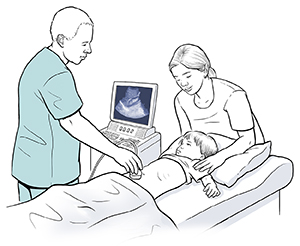
Your child will then lie down on an exam table. The lights may be dimmed in the room. This helps the radiologist see the images as they appear on a video screen.
-
The healthcare provider will put some warm gel on your child’s lower stomach, or pelvic area. The gel helps the sound waves travel through the body.
-
The healthcare provider will move a wand, or transducer, back and forth over the skin on your child’s pelvic area. As this is done, the transducer will emit sound waves. It will then pick up the sound waves as they echo or bounce back.
-
A computer will turn the sound waves into images. The images will appear on a video screen. They show the organs and other structures in real time.
-
At points during the test, the healthcare provider may ask your child to move into different positions or to hold their breath for a short time.
-
Once the test is done, the healthcare provider will wipe off the gel.
During the pelvic ultrasound, your child will need to stay still as much as possible. Moving around too much can distort the images. If you have a younger child, distracting them during the test may help. Try bringing along a stuffed animal or some other toy.
What happens after a pelvic ultrasound
Your child can go back to normal activities after the test. The radiologist may give you the results right away. Or you may get the results in a few days from your child’s healthcare provider.
Risks of a pelvic ultrasound
A pelvic ultrasound is a safe and painless test. It has no risks. It does not use radiation.
Online Medical Reviewer:
Irina Burd MD PhD
Date Last Reviewed:
2/1/2022
© 2000-2024 The StayWell Company, LLC. All rights reserved. This information is not intended as a substitute for professional medical care. Always follow your healthcare professional's instructions.