Chest Echocardiography (Transthoracic)
An echocardiogram (echo) is an imaging test that uses ultrasound to get pictures of your heart. It helps your healthcare provider look for symptoms that may be linked to how well your heart works. A transthoracic echo is sometimes called a TTE. It may also be called a surface echo. This is because the test is done outside your body. The pictures are taken from the outside (surface) of the chest wall. This test helps your provider assess your heart.
This test:
-
Is safe and generally painless
-
Can be done in a hospital, test center, or healthcare provider’s office
-
Bounces harmless sound waves (ultrasound) off the heart. It uses a handheld device (probe or transducer) that looks like a microphone.
-
May cause discomfort from the echo probe being pressed against the bony areas of the chest. This goes away once the probe is moved.
-
Lets your healthcare provider look at the size and shape of your heart. They can also see the size, thickness, and movement of your heart's walls, and the heart's pumping strength.
-
Shows if the heart valves are working correctly. It can also show if blood is leaking backward through your heart valves (regurgitation). Or if the heart valves are too narrow (stenosis).
-
Shows if you have a tumor or infectious growth around your heart valves
-
Will help your healthcare provider find out if there are problems with the outer lining of your heart (pericardium)
-
Shows problems with the large blood vessels that enter and leave the heart
-
Shows blood clots in the heart chambers
-
Shows abnormal holes between heart chambers
Before your echo
-
Discuss any questions or concerns you have with your healthcare provider.
-
Tell the healthcare provider about any over-the-counter or prescription medicines you're taking. This includes vitamins, herbs, or other supplements.
-
Allow extra time for checking in. Bring your insurance cards and ID.
-
Wear a 2-piece outfit for the test. You may be asked to remove clothing and jewelry from the waist up. If so, you’ll be given a short hospital gown.
-
An IV (intravenous) tube may be put into a vein in your arm or hand. This will be done if contrast dye or bubbles will be injected during the study.
During your echo
-
Small pads (electrodes) are placed on your chest to check your heartbeat.
-
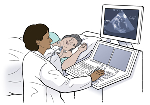
A probe coated with gel is moved firmly over your chest. The probe creates the sound waves that make pictures of your heart. If you're overweight, the technician may have to press down hard on the chest wall to get better pictures. This may be uncomfortable over bony areas. Tell your technician if you're uncomfortable.
-
At times, you may be asked to breathe out or hold your breath for a few seconds. Air in your lungs can affect the pictures.
-
The probe may also be used to do a Doppler study. This test measures the direction and speed of blood flowing through the heart. During the test, you may hear a whooshing sound. This is the sound of blood flowing through the heart.
-
The technician may use IV contrast dye. This helps to make the pictures clearer. Or they may use agitated saline. This helps to follow blood flow through the heart's chambers.
-
The pictures of your heart are stored electronically. This is so your healthcare provider can review them later.
 |
| During an echo, images of your heart appear on a monitor. |
Your test results
Your healthcare provider will discuss your test results with you during a future office visit. The test results help the healthcare provider plan your treatment and any other tests you need.
Online Medical Reviewer:
Ronald Karlin MD
Online Medical Reviewer:
Stacey Wojcik MBA BSN RN
Online Medical Reviewer:
Steven Kang MD
Date Last Reviewed:
4/1/2024
© 2000-2025 The StayWell Company, LLC. All rights reserved. This information is not intended as a substitute for professional medical care. Always follow your healthcare professional's instructions.